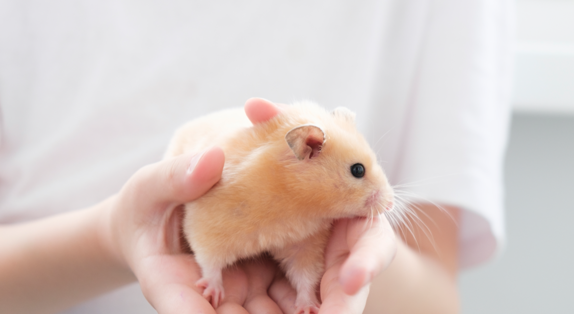
Exotic pets, including rabbits, hamsters, hedgehogs, reptiles, and birds, are small with delicate body structures, making a comprehensive clinical examination difficult. Imaging technologies such as X-rays and ultrasound play an important role in the diagnosis of exotic pets. This article will introduce the applications and procedures of imaging in exotic pet medicine, so owners can learn more about it.
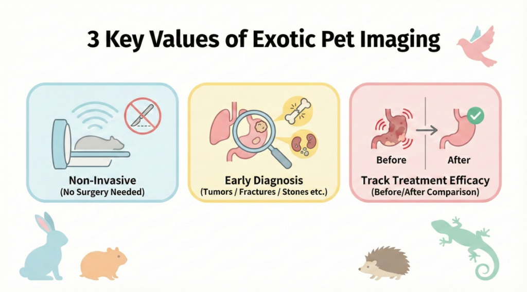
The Importance of Imaging
- Non-Invasive Examination: X-rays and ultrasound can provide a clear view of bones and internal organs without the need for surgery.
- Early Diagnosis: Imaging can help detect tumors, fractures, organ enlargement, and internal stones, helping with early treatment.
- Assessing Treatment Effectiveness: Imaging can be used to track changes in a condition after surgery or during treatment.
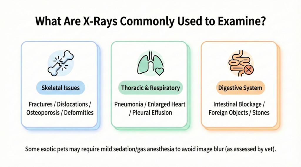
X-ray Applications
- Bone Problems: Such as fractures, dislocations, osteoporosis, and bone deformities.
- Respiratory Tract and Thorax: To evaluate pneumonia, an enlarged heart, or fluid accumulation in the chest.
- Digestive System: To check for intestinal blockages, foreign objects, or gallstones.
To prevent a blurry image caused by the animal moving, some exotic pets may need mild sedation or gas anesthesia during the X-ray.
Ultrasound Applications
- Abdominal Organs: To check the liver, kidneys, bladder, uterus, or ovaries, helping to find lumps, cysts, or inflammation.
- Cardiac Ultrasound: To evaluate heart valve function and blood flow.
- Reproductive System: To confirm pregnancy and fetal development, and to monitor for egg binding or difficult labor.
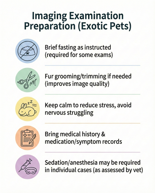
Safety and Preparation The amount of radiation used in imaging is extremely low and has limited effect on pets and owners. You must follow the vet’s instructions to fast the pet or prepare the hair before the examination to prevent the pet’s stomach or hair from affecting the image quality. Some reptiles or small animals may need mild anesthesia to prevent movement from blurring the image. Owners should discuss the risks of anesthesia with the vet.
Imaging Procedure The procedure includes making an appointment, preparing before the examination, calming the animal on the day of the examination, and waiting for the report. The vet will explain the report and recommend subsequent treatment. The cost of imaging varies depending on the equipment, the need for anesthesia, and the area being examined. If anesthesia or sedation is required, there will be an additional fee.
Frequently Asked Questions (FAQ)
- Will imaging harm my small animal? No. The amount of radiation from an X-ray is low, and an ultrasound is safe and harmless.
- Is anesthesia necessary for imaging? It depends on the individual animal. Active or nervous exotic pets may need sedation to get a clear image.
- Is fasting required for an imaging exam? Some exams require short-term fasting. Please follow your vet’s instructions.
- What should I be aware of after the exam? If the pet had anesthesia, it should stay at the clinic for observation. If not, it can go home to rest and follow the vet’s treatment recommendations.
- How often are imaging exams needed? It depends on the condition, but if there are abnormal symptoms or a need for post-surgery follow-up, imaging may be needed.
Cityvet Imaging Services
Cityvet is committed to providing outstanding comprehensive veterinary medical services, upholding the core values of “professionalism, care, and trust”. We aim to offer reasonably priced services, utilize advanced equipment, and provide detailed explanations of various treatment methods to owners, ensuring comprehensive medical care for every pet. To book a small animal imaging appointment, please call the Tsuen Wan clinic at 2623 5500 or the Yuen Long clinic at 2477 9990. Our addresses are: G/F, 186 Sha Tsui Road, Tsuen Wan / G/F, Fuk Shun Building, 25 Ping Wai Street, Yuen Long.
Disclaimer: This article provides general information only and is not a substitute for professional medical diagnosis. If you have any concerns, please schedule a veterinary appointment as soon as possible.